This is part 2 in a series about finding a solution to my chronic pain. Best to start with part 1 before reading this one.
Let’s do a quick refresher before I add more new information. Normally, the brain has very clear distinctions between body parts—this leg vs that one, right foot vs left, etc. With chronic pain those distinctions become muddied and the brain gets confused about where the painful body part starts and ends, how that body part is moving, and if it’s still safe. When the brain isn’t sure what’s happening and where, it requires more information to determine about whether or not you are in danger. To gather that information, it sends a signal that manifests as pain—anything from an itch, tingling or cramping to a sharp sensation that grabs your attention. This signal is magnified in individuals who’ve experienced (or are currently experiencing) one or more of the following: a recent surgery, pain in other parts of their body, and anxiety. Individuals experiencing those conditions often have a hypersensitive nervous system that is constantly on the lookout for threats of any kind. Given that I checked every one of these boxes, it’s no wonder I was dealing with chronic pain issues!
The process of Graded Motor Imagery (GMI) works to reset the brain’s body map by demonstrating to the brain that movement and touch are safe. And as the brain begins to see that the body is no longer in danger, it decreases the sensitivity of the nervous system accordingly. Fascinating, huh?
I hope that made sense because now I’m going to layer on some additional new science. To make these changes to the brain, we essentially need to de-emphasize the maladaptive pathways and reinforce the new ones. Scientists have found that the best way to do this is by targeting mirror neurons. They have a big influence on how safe or threatening a movement is to the brain. They’re also important because the information they gather is used to update the brain map.
So what is a mirror neuron, exactly? Well, mirror neurons are a type of brain cell that help us understand and relate to other people. They activate anytime we perform an action, witness someone else performing an action, or even *think* about moving one of our body parts. That’s why we often have intense responses even when something isn’t happening to us—think about watching an action or horror movie, or seeing someone get hurt. It’s like your brain is “mirroring” the other person’s actions or feelings, even though you’re not actually doing them yourself.
Mirror neurons help explain why pain can be influenced by what we see, think, and feel—not just what’s happening in the body. When you see someone else get hurt or look like they’re in pain, parts of your brain activate as if you might be experiencing that pain yourself. At their essence, mirror neurons help your brain simulate another person’s experience without you having to do the thing yourself.
Pain is a learned and reinforced response that happens in conjunction with mirror neurons. When a body part has been in pain a long time, the brain becomes very good at predicting pain. Mirror neurons contribute to this by reinforcing pain when the brain sees certain movements and reacting when it expects an activity to hurt (even if that part hasn’t moved yet). That’s why watching someone move or imagining yourself moving can trigger pain signals! The good news is that while mirror neurons often reinforce pain unintentionally, they can also help the brain unlearn it. That’s where GMI comes in.
And now, back to my story . . .
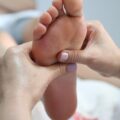
When I had my rights and lefts back in order, Jen had me do a visualization exercise where I looked at my injured foot while she gave me a series of prompts to do with my foot. “Imagine yourself sitting at your desk with your foot resting on the floor, now imagine yourself climbing the stairs, now step into high heels, now come down the stairs.” And so it went for a few minutes each session, followed by 20-30 minutes of therapeutic massage and foot manipulation/rotation.
I thought this imagining stuff sounded pretty useless, if I’m honest, but Jen gave me the example of high-level athletes or surgeons who religiously visualize each step in their process before a big event. The visualizations we did are part of step 2, or Motor Imagery, which is the process of imagining—but not doing—movement. Thanks to mirror neurons, neural pathways are activated just by imaging an activity. This allows the brain to practice safe movement without any physical strain happening to the injured body part. And each time this is done, the brain map gets updated with new information reinforcing that movement of that injured body part is normal and safe.
After practicing visualization foot exercises for a few weeks, I was surprised to realize that my foot was demanding less and less of my attention. It was mind-boggling that in six weeks of physical therapy we’d yet to do a traditional PT activity, but here I was experiencing symptom relief despite that.
Jen said I was ready to try the next step in the process. I assumed we’d be starting on foot-focused strengthening next, but nope, Jen surprised me yet again by pulling out a traditional door mirror, like the kind you had in your college dorm. She had me sit on a chair, remove my socks, and put the mirror—tall side up—between my knees with the mirrored side facing my left foot.
I had no idea what we were up to, and once again the process seemed rather bizarre, but since whatever we were doing together seemed to be working, I went along with it. With the mirror in place, we then proceeded to repeat the visualization exercises, except this time she had me look at the reflection of my foot in the mirror while doing it. Her next instruction was to roll my foot laterally then forward and backward, all while staring at it in the mirror. “Jen,” I grumbled, “This isn’t my hurt foot. How could this possibly be helping?” She then proceeded to explain step 3 to me, staying calm and cool as she confronted my growing frustration. “Sarah, what do you see when you look in the mirror?” The answer seemed a bit too obvious. “Uhhh, my left foot.” She nodded and continued, “But what does it look like?”. Then something clicked and a lightbulb turned on. “My right foot?” “Precisely,” she replied. “We are tricking your brain into thinking your right foot is moving without you actually having to move it. We’re going to let your brain watch as your ‘right foot’ moves safely until we convince it that the foot is better and doesn’t need protecting.” Now it was starting to make sense!
The next day, I faithfully did my foot-rolling homework in front of the mirror—morning, noon, and night. To my shock, I woke up the following day with pain that rivaled what I’d felt just two months after surgery. I was a bit shaken by the obvious setback. Thankfully, I had my second weekly PT session that day, and I made my displeasure very clear. “What is this witchcraft?” I half-jokingly exclaimed before relaying what had happened. “I’m in worse pain than when we started!” Jen looked excited though, which annoyed me. “Okay, we went too far,” she acknowledged. “I thought you weren’t ready for step 3, but the pain you’re feeling is feedback from your body that it was too much, too fast.” I couldn’t seem to let go of my annoyance. “This is ridiculous,” I scoffed, “how could rolling my non-injured foot possibly cause this much pain in my opposite foot?!”
Jen assured me that we were still progressing; that there were always backslides on the path to healing. So back I went to imagining foot movements instead of actually doing them. She was right though, because when we came back to mirror exercises two weeks later, my body didn’t have the same reaction. She viewed that as a major win and as a result began pushing me to do more of the activities I did pre-injury. “Take a hike. Walk barefoot in your house. Go back to the gym.” With her encouragement, I began revisiting old favorites and was delighted to find that I had a lot more tolerance for activity. For example, I could walk barefoot now, my feet could endure a two-hour square-dancing class with lots of foot impact, and I could walk outside on uneven surfaces. It was all rather exciting!
Each session I had more new wins to report and in early November she said, “It’s time to start strengthening your foot together—let’s go do some exercises.” FINALLY, I thought. About damn time! I started towards the weight area but she stopped me. “Hold up. Socks and shoes off please. All your strengthening work will be done barefoot.” Again, not what I was expecting, but I rolled with it. Over the next 6 weeks we did a series of barefoot exercises that worked on my balance, strengthened my toes’ ability to grip things, and conditioned my foot to be able to take load on the outer edge where my injury was. My foot, which had looked very emaciated and vein-y post surgery, now was filling out with muscle.
While my healing isn’t fully complete, I’m back to doing the things I love—with only mild, manageable pain. Even more meaningful, my foot no longer occupies my every thought. What once felt like it was running my life has faded into an occasional afterthought. I may not fully understand everything my brain, body, and nervous system learned through the work Jen and I did together, but I do know this: the changes I feel are real, and they’ve given me my life back.
**My eternal gratitude to Jen both for the remarkable care, but also for collaborating with me on this post to ensure all the science-y bits were accurate. I learned so much during this process and am pleased to be able to share that knowledge with a wider audience in the hopes that more people can find relief from chronic pain.**